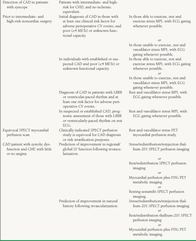
Guidelines suggest vasodilators as the first option 1. Pharmacologic stress for myocardial perfusion imaging fell out of my first experiment measuring coronary flow during progressive stenosis in 1972 published in 1974 the arteriogram and flowmeter dramatically showed the 3 fundamental physiological concepts underlying all stress myocardial perfusion imaging.

Dosing And Administration Protocol Lexiscan Regadenoson
Cv scan perfusion pharmacologic. A stress myocardial perfusion scan assesses blood flow to the heart muscle when it is stressed. A cardiac perfusion test tells your doctor if the muscles of your heart are getting enough blood. Its also known as myocardial perfusion imaging or a nuclear stress test. You might need this test if. Noninvasive imaging modalities are used to evaluate patients with or suspected of having cardiovascular cv disease. There are currently 3 vasodilators approved for myocardial perfusion stress testingdipyridamole adenosine and regadenoson.
Cardiovascular magnetic resonance cmr is a relatively unique imaging modality in that it can be used to depict left ventricular lv wall motion and myocardial perfusion with high spatial resolution in virtually any imaging plane without exposure to ionizing. With radionuclide myocardial perfusion imaging pharmacologic stress may be performed with an inotropic agent or vasodilator. Your heart rate and blood pressure will be monitored. The first following a period of rest and the second following a period of stress ie exercise. Pharmacologic myocardial perfusion scan. Instead you will lie on the table in the heart scanner while a medicine is injected into your iv.
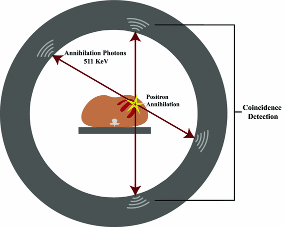
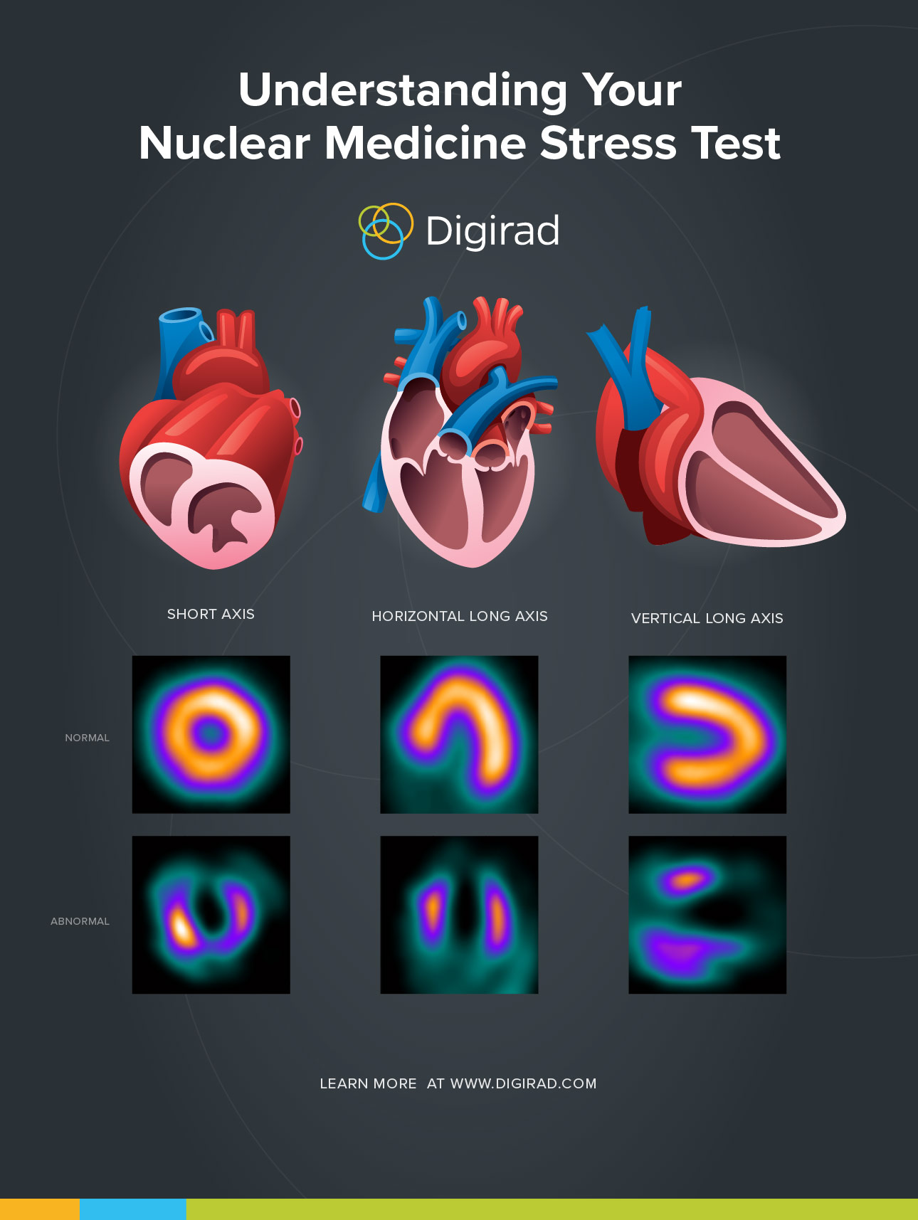
Started in 1995 this collection now contains 6806 interlinked topic pages divided into a tree of 31 specialty books and 736 chapters. The tracer will be injected into your iv line. After the radioactive tracer is injected a special type of camera is used that can detect the radioactive energy being released. A stressrest myocardial perfusion imaging mpi study is a type of stress test that uses pet or spect imaging of a patients heart before and after exercise to determine the effect of physical stress on the flow of blood through the coronary arteries and the heart muscle. It can show areas of the heart muscle that arent getting enough blood flow. Myocardial perfusion imaging mpi is a non invasive imaging test that shows how well blood flows through perfuses your heart muscle.
You will not exercise on a treadmill. A myocardial perfusion spect single photon emission computed tomography study also called a cardiac stress rest test helps your doctor evaluate your hearts blood supply. Two sets of images showing blood flow are obtained. The heart can be stressed with exercise or if unable to exercise a medication can be used.


